Vol 66, No 3 (2021)
- Year: 2021
- Published: 10.07.2021
- Articles: 8
- URL: https://virusjour.crie.ru/jour/issue/view/50
Full Issue
ANNIVERSARY DATES
 173-181


ORIGINAL RESEARCHES
Markers of viral hepatitis E (Hepeviridae, Orthohepevirus, Orthohepevirus A) in the imported Old World monkeys
Abstract
Introduction. Viral hepatitis E is a zooanthroponotic disease that occurs in humans and various animals, including monkeys. It is caused by hepatitis E virus (HEV) (Hepeviridae, Orthohepevirus: Orthohepevirus A), for which 8 genotypes have been described to date. Among them, strains of genotypes 1 and 2 have been isolated from humans, strains of genotypes 3 and 4 from humans and animals, and strains of genotypes 5–8 from animals only. The main threat of the disease is associated with the documented zoonotic transmission of HEV genotypes 3, 4, 7, and 8, to humans through infected meat, blood and milk. Thus, monkeys could be involved in the transmission of HEV.
The aim of this work was to study serological and molecular genetic markers of HEV infection in strepsirrhines (Old World monkeys, Cercopithecoidea), imported to the Adler Primate Center from various regions of the world (Tanzania, Vietnam, Mauritius).
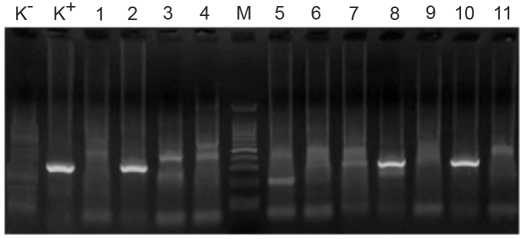
Material and methods. Fecal (n = 224) and blood serum samples (n = 395) from cynomolgus (Macaca fascicularis) and vervet monkeys (Chlorocebus pygerythrus) were examined by the enzyme-linked immunosorbent assay (ELISA) and reverse transcription polymerase chain reaction (RT-PCR).
Results and discussion. The data obtained show the high detection rate (51.8%) of IgG antibodies to HEV among 5 groups of cynomolgus monkeys imported from Vietnam, with a predominance of highly reactive sera (84%). High detection rate of IgM antibodies in these animals (10.4%) was observed, with the large number of IgM-reactive sera in one particular group of animals (36.8%). The fact of detection of HEV RNA in two groups of cynomolgus monkeys (11.9% and 5.7%) is of particular importance. All HEV sequences of isolated from monkeys belonged to genotype 4.
Conclusion. Our data indicate that monkeys (in particular, cynomolgus monkeys) can serve as a natural reservoir of HEV genotype 4 for humans. This requires an appropriate set of anti-epidemic measures in a number of situations.
 182-188


Monoclonal antibodies to hemagglutinin of influenza A/H7N3 virus (Orthomyxoviridae: Alphainfluenzavirus: Influenza A virus)
Abstract
Introduction. Variants of influenza virus A/H7 have the same high pandemic potential as A/H5. However, the information about the antigenic structure of H7 hemagglutinin (НА) is considerably inferior in quantitative terms to similar data for H5 НА.
The aims of the study were development and characterization of the monoclonal antibodies (MAbs) panel for HA subtype H7 of the influenza A virus.
Material and methods. Viruses were accumulated in 10-day-old chicken embryos. Purification and concentration of the virus, determination of protein concentration, preparation of MAbs and ascitic fluids, hemagglutination and hemagglutination inhibition (HI) tests, assessment of antibodies’ activity in indirect enzyme-linked immunosorbent assay (ELISA), as well as determination of MAbs isotypes and neutralization reaction (NR) were carried out by standard methods.
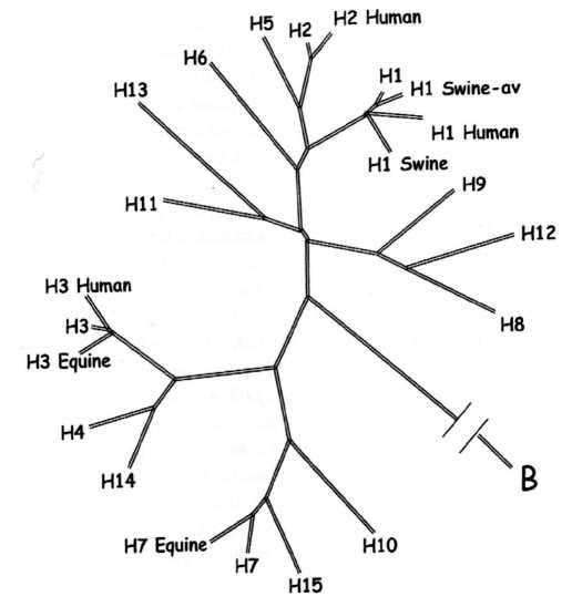
Results. The obtained MAbs to А/mallard/Netherlands/12/2000 (H7N3) strain were studied in HI test with a set of strains of different years of isolation belonging to different evolutionary groups. MAbs had a reduced reactivity compared to the immunogen-virus for all the studied strains. Cross-interaction of MAbs 9E11 and 9G12 in HI test with influenza A/H15 virus has been observed.
Discussion. Influenza A agent with H7 HA variant could serve as a potential cause of a future pandemic. Development of the MAbs panel for subtype H7 HA is an urgent task for both veterinary medicine and public health.
Conclusion. The obtained MAbs can be used not only for epitope mapping of the H7 HA molecule (currently insufficiently studied) and as reagents for diagnostic assays, but also for determining common («universal») epitopes in HA of different strains of this subtype.
 189-197


Modulation of endothelial factors activity in human endothelial cells in influenza A(H1N1)pdm09 virus infection
Abstract
Introduction. Influenza A virus infection can lead to endothelial dysfunction (ED), including apoptosis of endothelial cells and modulation of endothelial factor activities. Affected biochemical factors may include those playing important roles in vascular homeostasis. However, the effect of this pathogen on the expression pattern of key endothelial factors is still unknown.
The aim of this work was to study the expression of endothelial nitric oxide synthase (eNOS) and plasminogen activator inhibitor-1 (PAI-1, serpin E1) in the EA.hy926 endothelial cells. Research objectives: to assess expression of eNOS and PAI-1 in endothelial cells infected with influenza virus A(H1N1)pdm09, and to identify homologous fragments in structure of viral proteins and endothelial factors.
Material and methods. Cells were infected with influenza virus A/St. Petersburg/48/16 (H1N1)pdm09 and analyzed in dynamics in 6, 12, 18, 24, 48, and 72 hrs post infection (hpi). Detection of endothelial factors expression levels was performed by immunocytochemical method (ICC) using antibodies for eNOS and PAI-1 while quantitative assessment of expression levels was carried out by program Nis-Elements F3.2 («Nikon», Japan). The search for homologous sequences between viral proteins and eNOS and PAI-1 was performed by computer comparison. Sequences were analyzed as fragments 12 amino acid residues (aar) in length.
Results and discussion. eNOS expression in infected cells had decreased to 7.9% by 6 hpi (control was taken as 100%) to 3.3% at 72 hpi. PAI-1 expression varied significantly over the course of the experiment: by 6 hpi it had decreased to 49.6%, and to 43.2% by 12 hpi. Later PAI-1 levels were: 116.3% (18 hpi); 18.9% (24 hpi); 23.5% (48 hpi), and 35% (72 hpi).
Conclusion. These results indicate that influenza A infection of endothelial cells causes a significant decrease in eNOS expression, while modulating PAI-1 one. The described phenomenon can be used in the further development of directions of pathogenetic therapy of vascular complications of infection caused by this pathogen.
 198-210


Changes in the genome of African swine fever virus (Asfarviridae: Asfivirus: African swine fever virus) associated with adaptation to reproduction in continuous cell culture
Abstract
Introduction. African swine fever virus (ASFV) is a large, double-stranded DNA virus in the Asfarviridae family. It is the causative agent of African swine fever (ASF). Only the genome of BA71V strain, adapted to Vero cell culture, was fully analyzed.
The aim of this study was analyzing the complete genome sequence of two strains of adapted to the growth in CV-1 cell culture (CC) ASFV obtained after 30 and 50 passages, in comparison to the parental virus.
Material and methods. ASFV isolate Odintsovo 02/14 (parental), ASFV adapted variants ASFV/ARRIAH/CV-1/30 and ASFV/ARRIAH/CV-1/50 were all used to extract genomic DNA (gDNA). Sequencing library was constructed using the «Nextera XT DNA library preparation kit» («Illumina», USA).
Results. Genomes of ASFV/ARRIAH/CV-1/30 and ASFV/ARRIAH/CV-1/50 consisted of 186 529 bp and 186 525 bp, respectively. Total 78 single nucleotide polymorphisms (SNPs) were identified between the parental Odintsovo 02/14 and the two high passaged strains, as well as a 2947 bp large-size deletion in the 3’ variable region of adapted viruses was detected.
Discussion. ASFV as a DNA-containing virus may not have a very high level of mutation, but this is the second study showing that adaptation to growth in continuous CC leads to large deletions in the genome of the virus.
Conclusion. Mutations in the protein-coding regions of the genome can be synonymous and non-synonymous, i.e. leading to amino acid substitution. Additional research is needed to understand the influence of the mutations described in the adaptation process on the reproduction of the virus and its virulence.
 211-216


Prevalence of human papillomavirus (Papillomaviridae; Human papillomavirus) of high carcinogenic risk based on the results of screening of three anatomical loci in men stratified by sexual behavior and HIV status
Abstract
Introduction. Human papillomavirus (HPV) of high carcinogenic risk (HCR), in addition to being the etiological agent of cervical cancer, also contribute to development of cancer of the anus, vagina, penis, vulva and oropharyngeal cancer. In this connection, further study of the biological properties of this agent and its prevalence in different populations is an urgent task.
The aim of the study was to examine the prevalence of HCR HPV in three anatomical loci in men stratified by HIV (human immunodeficiency virus) infection status (negative, HIV+/positive/HIV–) as well as by sexual behavior: men who have sex with men (MSM), heterosexual men (HM).
Material and methods. The study included 256 men from Moscow and Moscow region: 73 МSМ/HIV+, 66 МSМ/ HIV–, 58 HM/HIV+, and 59 HM/HIV–. All men were tested for 14 HCR genotypes of HPV (16, 18, 31, 33, 35, 39, 45, 51, 52, 56, 58, 59, 66, and 68). Smears were taken from three anatomical loci: urethra, anus, oropharynx. Testing was preformed using real-time polymerase chain reaction assay (PCR-RT).
Results. The highest prevalence of HCR HPV detection, regardless of the locus, was recorded for МSМ/HIV+ (82.2%), and the lowest for HM/HIV (20.3%). The highest detection of HCR HPV in scrapings of epithelial cells from anus was recorded for МSМ/HIV+ (79.5%). The highest incidence of this pathogen in oropharynx also was registered for МSМ/HIV+ (13.7%). The highest incidence of HCR HPV in scrapings of epithelial cells from urethra was recorded for HM/HIV+ (24%). The prevalence of HCR HPV among men was found to differ markedly depending on the anatomical locus, HIV status and sexual behavior.
Discussion. For the first time, there were obtained data on the prevalence of HCR HPV in men with different patterns of sexual behavior and HIV status in the Moscow region.
Conclusion. Screening for HCR HPV in male population based on the identification of 14 genotypes of the virus in three anatomical loci (urethra, oropharynx, anus) by PCR-RT will provide the information necessary to improve the system of epidemiological monitoring and proper planning of preventive measures among men with any risk factors for HPV persistence (presence of HIV infection and/or belonging to the MSM group). HPV screening algorithm development is required for men considering their HIV status and sexual behavior. We recommend testing for 14 HCR HPV genotypes in three loci (urethra, anus, oropharynx).
 217-226


Expression of integrins β1, α4 and cell adhesion molecule ICAM-1 in the presence of sodium deoxyribonucleate with ferrum complex (DNA-Na-Fe) by MT-4 cells transformed by human T-lymphotropic virus type 1 (Retroviridae: Orthoretrovirinae: Deltaretrovirus: Human T-lymphotropic virus type 1)
Abstract
Introduction. The important role of integrins (IG) in the initiation and development of cancer processes makes these structures convenient targets for the development of immunomodulatory therapeutic drugs that have an effect directly on these molecules. Among the latter, IG β1, α4 and cell adhesion receptor ICAM-1 (intercellular adhesion molecule 1) are of particular interest. Immunomodulators are capable of changing the IG activity through non-specific mechanisms, which, however, in some cases can cause a decrease in the protective functions of the immune system and health deterioration.
The aim of the study was to determine the effect on the levels of cellular expression and the nature of IG metabolism of the drug sodium deoxyribonucleate with ferrum complex, DNA-Na-Fe, which is having been used in the Russian Federation as an immunomodulatory agent, but whose action has not been studied in details so far.
Material and methods. We used 2 variants of the neoplastic CD4+ T-lymphocyte cell line transformed with human T-lymphotropic virus type 1 (HTLV-1) of the Retroviridae family, MT-4 (MT-4/1 and MT-4/2). The indicated variants were characterized by different levels of expression of the protein activation markers CD28 and CD38. After cell culture in the presence of 500 μg/ml DNA-Na-Fe, the expression levels of IG β1 (CD29), α4 (CD49d), and ICAM-1 (CD54) were studied by flow cytometry.
Results. The cells of the both lines contained many membrane proteins CD29+ (90.4% ± 4.5) and CD54+ (97.9% ± 1.4), while small percentage of cells contained protein CD49d+ (1.9% ± 1.0). No changes in the expression of the studied proteins were observed in the presence of the drug.
Discussion. The levels of IG β1, α4 and ICAM-1 expression may serve as one of the phenotypic characteristics of MT-4 cells. The obtained data are of great importance because the peculiarities of CD4+ T-lymphocytes transformation and their metabolism during HTLV-1 infection have not been sufficiently studied so far.
Conclusion. The results of this work may be helpful in determining the pathogenesis of HTLV-1-induced diseases, some types of malignancies, and in searching for new specific pharmacological agents, including molecularly targeted ones. The results of the study will help to expand the existing knowledge on the markers of MT-4 cell line.
 227-232


OBITUARY
 233-234


