


Determination of cold-adapted influenza virus (Orthomyxoviridae: Alphainfluenzavirus) polymerase activity by the minigenome method with a fluorescent protein
- Authors: Ivanov P.A.1, Lyashko A.V.1, Kost V.Y.2, Lomakina N.F.1, Rtishchev A.A.3, Bunkova N.I.1, Timofeeva T.A.1, Balanova M.A.1, Ionov S.A.1,4, Gorikov D.V.1,4, Markushin S.G.3
-
Affiliations:
- N.F. Gamaleya National Research Centre for Epidemiology and Microbiology, the Russian Ministry of Health
- Shemyakin-Ovchinnikov Institute of Bioorganic Chemistry of the Russian Academy of Sciences
- Mechnikov Research Institute for Vaccines and Sera
- Mendeleev University of Chemical Technology
- Issue: Vol 68, No 6 (2023)
- Pages: 526-535
- Section: ORIGINAL RESEARCHES
- URL: https://virusjour.crie.ru/jour/article/view/16588
- DOI: https://doi.org/10.36233/0507-4088-203
- EDN: https://elibrary.ru/lxvxzf
- ID: 16588

Cite item

Abstract
Introduction. Polymerase proteins PB1 and PB2 determine the cold-adapted phenotype of the influenza virus A/Krasnodar/101/35/59 (H2N2), as was shown earlier.
Objective. The development of the reporter construct to determine the activity of viral polymerase at 33 and 37 °C using the minigenome method.
Materials and methods. Co-transfection of Cos-1 cells with pHW2000 plasmids expressing viral polymerase proteins PB1, PB2, PA, NP (minigenome) and reporter construct.
Results. Based on segment 8, two reporter constructs were created that contain a direct or inverted NS1-GFP-NS2 sequence for the expression of NS2 and NS1 proteins translationally fused with green fluorescent protein (GFP), which allowed the evaluation the transcriptional and/or replicative activity of viral polymerase.
Conclusion. Polymerase of virus A/Krasnodar/101/35/59 (H2N2) has higher replicative and transcriptional activity at 33 °C than at 37 °C. Its transcriptional activity is more temperature-dependent than its replicative activity. The replicative and transcriptional activity of polymerase A/Puerto Rico/8/34 virus (H1N1, Mount Sinai variant) have no significant differences and do not depend on temperature.
Full Text
Introduction
Influenza A virus, a member of the Orthomyxoviridae family, has a segmented single-stranded negative-sense RNA genome. Each of the 8 segments encodes 1–2 or more viral proteins and is packaged as a ribonucleoprotein (RNP). The latter includes viral negative-strand RNA (viral genomic RNA, vRNA), covered with molecules of viral protein NP in a bead-like manner, and one copy of viral polymerase in the form of a heterotrimer of proteins PB1, PB2, PA. From this structure in the nucleus of an infected cell, viral polymerase carries out transcription and subsequent replication of viral RNA. Segment 8 encodes two proteins, NS1 and NEP/NS2, the latter being translated from spliced messenger RNA (mRNA) and participating in the transport of newly formed RNPs from the nucleus to the cell membrane for the assembly of new virions. Segments 4 and 6 encode surface glycoproteins HA and NA, which serve as the main antigenic determinants provoking the immune response in the infected organism [1]. HA ensures interaction of the virion with receptors and penetration of the viral genome into the cell, while NA enables the egress of viral progeny from the cell.
The influenza virus has been a problem for mankind for centuries and is most prevalent during seasonal influenza epidemics, which in some cases are characterized by high mortality. Vaccination remains one of the most effective preventive measures against influenza. Nowadays, there is a huge variety of influenza vaccines based on different technologies [1]. Among whole-virion vaccines, which include inactivated and live attenuated vaccines, the latter are considered the most effective. The mechanism of their action is based on the fact that the vaccine strain is able to replicate at a reduced temperature (25–33 °C), which corresponds to the temperature in the human nasal passages. In the lower respiratory tract, where the temperature exceeds 37 °C, the virus loses its ability to replicate and is eliminated from the body without causing disease. Contact with the vaccine virus activates humoral and cellular immunity, which protects the body from severe disease when it encounters a wild-type virus. At a certain point in time, attenuated cold-adapted strains A/Leningrad/134/17/57 in Russia and A/Ann Arbor/6/60 in the USA were used as live vaccines against influenza A H2N2 viruses [2–4]. Later, these strains were used as attenuation donors to obtain reassortant vaccine strains in which the genes of HA and NA surface proteins were replaced with genes of circulating actual strains [5–7].
Significant progress in the development of influenza vaccines has been achieved due to reverse genetics. The essence of this method is that each of the eight segments of the influenza A virus genome is cloned in a plasmid. From these plasmids, acting like a constructor, it is possible to assemble a virus with specific properties [8–10].
One of the viruses reconstructed by plasmid technology was the A/Puerto Rico/8/34 (H1N1) virus with high reproductive capacity in chicken embryos (CE). It was supposed to be used as an internal protein gene donor in the development of inactivated recombinant vaccines, in particular, against the H5N1 virus [11].
The use of reverse genetics in the production of influenza vaccines, including the live FluMist seasonal influenza vaccine (http://www.flu.org.cn/en/news-11930.html), is now license-approved in the USA [5]
Reverse genetics is widely used to study the function of viral proteins and the mechanisms of their interaction with host cellular factors. It is also used to elucidate the role of specific genome regions and point mutations in phenotype changes.
The use of plasmid technology makes it possible to conduct research without assembling an entire virus. For example, to determine the activity of viral polymerase, it is sufficient to assemble a construct of plasmids expressing PB1, PB2, PA, and NP proteins, also known as the minigenome. To assess the expression of the minigenome in transfected cells by fluorescent or luminescent luminescence, a reporter gene expressing green fluorescent protein (GFP) or luciferase is additionally added into the construct [12, 13].
Improvement of reverse genetics technology as applied to influenza virus consists of the design of plasmids for specific purposes, simplification of synthesis of viral gene copies in the form of cDNA by polymerase chain reaction (PCR) and the method of their incorporation into plasmids for cloning. The first systems for complete virus assembly included 12 plasmids, later the number of plasmids was reduced to 8 or less. The design of the double-stranded plasmid pHW2000 with two promoters made it possible to synthesize mRNA for viral protein translation from an inserted DNA fragment on one strand, and in the opposite direction, on the complementary strand, to obtain vRNA, which is subsequently incorporated into the RNP of mature virions. In this case, a strong cytomegalovirus (CMV) promoter, which is recognized by cellular RNA polymerase II, is used for mRNA synthesis; a cellular promoter for RNA polymerase I is used for vRNA synthesis [9, 10].
Studies at the molecular level of cold-adapted strains have shown that most of the mutations responsible for the ts-, ca-, att1-phenotype are localized in the genes of the polymerase complex. Thus, for strain A/Ann Arbor/6/60, point mutations in PB2 (N265S), PB1 (K391E, D581G, and A661T), and NP (D34G) were detected [14, 15]; for strain A/Leningrad/134/57, mutations were found in PB2 (V478L), PB1 (K265N, V591I), and NEP (M100I) [16]. Incorporation of these mutations into the corresponding segments of different influenza virus strains by reverse genetics resulted in the manifestation of ts-, ca-, and att-phenotype traits to different degrees depending on the virus strain in which the mutations were introduced. One of the possible mechanisms of attenuation and cold adaptation may be related to the impairment of transcriptional and/or replicative polymerase activity at elevated temperature. L. Rodriguez et al. carried out a study [15] where the minigenome method was used and it was shown that incorporating appropriate mutations into the polymerase segments of the A/Puerto Rico/8/34 (H1N1) strain resulted in a significant decrease in its activity when the temperature was increased to 37 and 39 °C.
During the search for new attenuation donors in Russia, a cold-adapted strain A/Krasnodar/101/35/59 (H2N2) was obtained, which differed from its wild-type progenitor by mutations in all segments except for segment 8 [17, 18]. Two single mutations were present in the polymerase proteins PB1 (I147T) and PB2 (V290L). To elucidate their role in ts-phenotype formation, similar substitutions were incorporated into A/WSN/33 (H1N1) virus by reverse genetics, resulting in virus variants that differed from the original strain by a single substitution in PB1 (I147T) or in PB2 (V290L). The mutant variants multiplied well in CE at 34 °C and poorly at elevated temperatures [19–21].
The objective of the present study was to master the influenza virus minigenome techniques and to create reporter constructs carring fluorescent protein in order to use them for studying the function of viral polymerases in the processes of transcription and replication at different temperature incubation. Polymerase protein (PB1, PB2, PA) and NP genes from the cold-adapted A/Krasnodar/101/35/59 (H2N2) strain and A/Puerto Rico/8/34 (H1N1, Mount Sinai variant) strain were taken for comparison.
Materials and methods
Cloning of PB1, PB2, PA, and NP genes
Human influenza virus A/Puerto Rico/8/34 (H1N1), Mount Sinai variant, was obtained from the collection of the D.I. Ivanovsky Research Institute of Virology.
Viral RNA was isolated from the allantois fluid of infected CEs using the QIAamp Viral RNA mini kit (#52904; Qiagen, Germany) according to the manufacturer’s instructions. Reverse transcription was performed at 42 °C for 1 h in 25 μl of a reaction mixture containing 8 μl of RNA, 1 μl of uni12 primer with a concentration of 50 ng/μl (13.5 nM), 10 μl of water, 1 μl of 10 mM dNTP, 5 μl of 5× buffer, and 100 units of MMLV (Alpha Enzyme LLC, Moscow). The obtained 3 μl of cDNA was used in 30 μl of PCR (94 °C – 15 s, 52 °C – 15 s, 72 °C – 1 min, 30 cycles) with specific primers to synthesize full-length genome segments [22] in the presence of 0.5 μl of Pfu enzyme (Alpha Enzyme LLC, Moscow). Amplified fragments were separated by electrophoresis in a 1.2% agarose gel containing ethidium bromide and eluted from the gel with Diatom DNA Elution kit (#D1031; Isogen Laboratories LLC, Russia). The purified segments were cloned into the pHW2000 vector by ligase-free method using T4 DNA polymerase [23]. The vector for cloning was kindly provided by Dr R. Webster (St. Jude Children’s Research Hospital, Memphis, USA).
Constructs of 8 phW2000 plasmids including genome segments for assembly of A/Krasnodar/101/35/59 (H2N2) virus were created in the laboratory of genetics of RNA-containing viruses of the I.I. Mechnikov Research Institute of Vaccines and Serums (Moscow) [24], as described previously [19, 20].
A reporter construct with the GFP gene
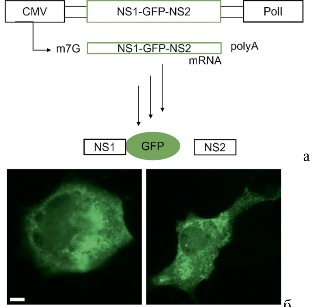
Segment 8 of A/Krasnodar/101/35/59 (H2N2) virus encoding NS1 and NEP/NS2 proteins (as a result of splicing) was replaced with a reporter construct created as described in [25]. The splicing site for NS2 was removed from this segment by PCR and ligase-free cloning methods [23]. The sequence of the segment between the NS1 stop codon and the 3`UTR was then replaced with a sequence containing the GFP gene and the spliced NS2 gene, separated by the sequence for peptide 2A, where the ribosome frameshift occurs. The final construct included NS1, GFP, the sequence for peptide 2A and the NS2 gene arranged sequentially in the same reading frame. Translation of the mRNA of this construct produced two distinct proteins, NS1, translationally fused to GFP, and NS2. The construct was named NS1GFPNS2 (Fig. 1 a). The CMV promoter was then removed from the vector part of this construct using PCR and ligase-free cloning techniques [23], and the construct was named dCMV (Fig. 2). Similarly, a plasmid construct in which the sequence of the NS1GFPNS2 segment was inverted was obtained. The construct was named dCMVrev (Fig. 3).

Fig. 1. NS1GFPNS2 reporter construct (control).
a – modified segment 8 of A/Krasnodar/101/35/59 (H2N2) influenza virus is inserted in the plasmid which includes the NS1 gene (with a removed splicing site for NS2), the green fluorescent protein gene (GFP), and spliced NS2 gene. Cellular RNA polymerase II uses the CMV promoter of the NS1GFPNS2 reporter to synthesize mRNA (mRNA) followed by translation of NS2 and fused NS1-GFP proteins; b – expression of the fused NS1-GFP protein in transfected Cos-1 cells cultured at 33 °C (left) and 37 °C (right). The scale ruler is 10 microns.
Рис. 1. Репортерная конструкция NS1GFPNS2 (контроль).
а – встроенный в плазмиду модифицированный сегмент 8 вируса гриппа А/Краснодар/101/35/59 (H2N2) включает ген NS1 (с удаленным сайтом сплайсинга для NS2), ген зеленого флуоресцирующего белка (GFP), сплайсированный ген NS2. Клеточная РНК-полимераза II с промотора CMV синтезирует мPHK (mRNA) конструкции NS1GFPNS2, с которой транслируются белки NS2 и NS1, трансляционно слитный с GFP; б – экспрессия слитного белка NS1-GFP в трансфецированных клетках Cos-1, культивируемых при 33 °C (слева) и 37 °C (справа). Масштабная линейка – 10 мкм.

Fig. 2. The dCMV reporter construct for evaluating the transcriptional activity of viral RNA-dependent RNA polymerase (RdRp).
The CMV promoter has been removed from a plasmid carrying inserted NS1GFPNS2 sequence. On one chain of the inserted double-stranded DNA fragment, cellular RNA polymerase I (PolI) synthesizes virionic RNA (vRNA) from the PolI promoter. Then, the viral RNA polymerase RdRp, expressed by the minigenome, uses this vRNA for synthesis of mRNA that translates NS1-GFP and NS2 proteins.
Рис. 2. Репортерная конструкция dCMV для оценки транскрипционной активности вирусной РНК-зависимой РНК-полимеразы (RdRp).
В плазмиде с встроенной последовательностью NS1GFPNS2 удален промотор CMV. На одной цепи встроенного двухцепочечного фрагмента ДНК клеточная РНК-полимераза I (PolI) с промотора PolI синтезирует вирионную РНК (vRNA), с которой вирусная РНК-полимераза RdRp, экспрессируемая минигеномом, в результате транскрипции синтезирует mRNA для трансляции белков NS1-GFP и NS2.

Fig. 3. The dCMVrev reporter construct for evaluating the replicative and transcriptional activity of viral RNA-dependent RNA polymerase (RdRp).
An inverted double-stranded DNA fragment NS1GFPNS2 is inserted in the plasmid in which the CMV promoter is deleted. Cellular RNA polymerase I (PolI) synthesizes a complementary NS1GFPNS2 chain (cRNA) of an inserted construct. Viral RNA-dependent RNA polymerase (RdRp) replicates viral RNA (vRNA) from cRNA. Then RdRp uses vRNA as a template for synthesis of mRNA for following translation of NS1-GFP and NS2 proteins.
Рис. 3. Репортерная конструкция dCMVrev для оценки репликативной и транскрипционной активности вирусной РНК-зависимой РНК-полимеразы (RdRp).
В плазмиду встроен инвертированный двухцепочечный ДНК-фрагмент NS1GFPNS2 и удален промотор CMV. Клеточная РНК-полимераза I (PolI) синтезирует комплементарную цепь NS1GFPNS2 встроенной конструкции (cRNA). Вирусная РНК-зависимая РНК-полимераза (RdRp) реплицирует с нее вирусную РНК (vRNA), на которой затем синтезирует mRNA для трансляции белков NS1-GFP и NS2.
The structure of the primers used in this study is available upon request. The structure of all constructs incorporated into plasmids was confirmed by sequencing.
Assessment of influenza virus minigenome activity by NS1-GFP protein expression
The immortal cell line Cos-1 was cultured at 37 °C in an atmosphere of 5% CO2 in DMEM medium containing 10% fetal calf serum and antibiotics. Cells were overseeded once every 2 days using 0.25% trypsin in Versen solution, and their confluency of 70–90% was maintained.For transfection, cells were seeded with a confluency of 70% in 6-well plates with coverslips at the bottom. Transfection was performed using Lipofectamine-3000 (Thermo Fisher Scientific, USA) according to the manufacturer’s instructions. 2 μg of DNA was added to a well of a 6-well plate. After transfection, cells were incubated overnight at 33 or 37 °C and fixed with 3% paraformaldehyde solution on phosphate buffer (PBS). Next, coverslips with attached fixed cells were washed with PBS and mounted microscopically using a solution containing 9.1% Moviol 4-88 and 2.3% glycerol in 100 mM Tris-HCl, pH 8.5.
To observe fluorescence in cells, an Imager M2 microscope (Carl Zeiss, Germany) with a FITC filter and a 40× objective lens was used. Cell images were acquired using an AxioCam 503 mono digital camera and saved in tiff format with 16-bit resolution. Images were processed and analyzed using ImageJ2 software. Before analyzing the image, background fluorescence was removed from the image. For this purpose, the average value of fluorescence in the area where cells were absent was measured in the image. The obtained value was subtracted from the fluorescence values for each point of the image. To exclude parts of the image outside of cells from the analysis, the brightness threshold was manually adjusted. Individual cells in the image were identified using the command to analyze particles. The average fluorescence level was then measured for each cell. Three independent transfection experiments were performed for each reporter construct. Between 10 and 40 measurements were taken in each experiment. The total number of measurements in all independent experiments for each construct was used to calculate the average fluorescence value.
Statistical analysis of data was performed using Statistica software (StatSoft, USA). The Mann–Whitney test was used for statistical analysis of differences in the fluorescence level at different incubation temperatures of transfected cells. Differences were considered statistically significant at p < 0.05.
Results and discussion
Minigenome and reporter construct with fluorescent protein
The minimal set of plasmids capable of replicating the function of influenza virus polymerase in an infected cell consists of four plasmids with PB1, PB2, PA, and NP segments. One chain of each plasmid vector contains CMV promoter, which is recognized by cellular RNA polymerase II synthesizing the mRNA from the inserted segment for subsequent translation of the viral protein. The opposite strand contains the promoter for cellular RNA polymerase I, which can synthesize vRNA from the same segment (Fig. 1 a).
In order to observe the functioning of such an incomplete genome (minigenome) in a living cell under the microscope with the help of fluorescence, a reporter construct was created. The control of the experiment was a plasmid with a modified segment 8, from which the NEP/NS2 protein and the NS1 protein with attached GFP were expressed separately. When cells were transfected with that single plasmid, green luminescence was observed in them (Fig. 1 b).
Transcriptional activity
The dCMV reporter plasmid (Fig. 2) with a deleted CMV promoter for cellular RNA polymerase II can function in a living cell only in the presence of viral RNA-dependent RNA polymerase (RdRp) generated by co-transfection of the dCMV reporter plasmid together with plasmids carrying the viral genes PB2, PB1, PA, and NP. Through cellular PolI, a vRNA(−) is synthesised from one DNA(+) strand of the integrated double-stranded dCMV reporter construct, on which the viral RdRp builds an mRNA for translation of the fluorescent protein. The presence of fluorescence indicates the transcriptional activity of the viral polymerase.
Replicative and transcriptional activity
In the case of the dCMVrev reporter plasmid (Fig. 3), in which the inserted NS1GFPNS2 fragment is inverted, a complementary cRNA(+) is synthesised in the cell due to the activity of cellular RNA polymerase I from the complementary DNA(−) chain of the insertion. Then replication takes place, i.e. as a result of activity of viral RNA polymerase vRNA(−) is formed, which at the stage of transcription serves as a template for synthesis of mRNA, from which the fluorescent protein is translated. Thus, the expression of NS1-GFP protein can be used to assess the replicative and transcriptional activity of the viral polymerase RdRp.
Activity of viral polymerase at different temperatures
The minigenome method was used to study the polymerase activity of A/Puerto Rico/8/34 (H1N1) and A/Krasnodar/101/35/59 (H2N2) viruses at different temperatures.
For each virus, a minigenome was obtained in Cos-1 cell culture by simultaneous transfection with plasmids expressing PB2, PB1, PA, and NP proteins together with a dCMV or dCMVrev reporter construct. The transfected cells were cultured at 33 or 37 °C. The transfection had an efficiency of 5–10%.
When cells were transfected with reporter constructs alone (without viral minigenome), specific GFP fluorescence was absent (data not shown). When reporter constructs were co-transfected together with plasmids carrying PB2, PB1, PA, and NP genes of both viruses, fluorescent luminescence was observed in transfected cells. In the case of the A/Puerto Rico/8/34 (H1N1) virus minigenome, there was no significant difference in the fluorescence of cells cultured at different temperatures (Fig. 4, 5, top row). However, when co-expressed with polymerase reporter constructs of cold-adapted A/Krasnodar/101/35/59 (H2N2) strain, cell fluorescence was significantly higher at a lower temperature (33 °C) (Fig. 4, 5, bottom row).

Fig. 4. Expression of NS1-GFP in Cos-1 cells after transfection with a reporter construct without a CMV promoter (dCMV) together with the minigenome of A/Krasnodar/101/35/59 (H2N2) virus (bottom row), or A/Puerto Rico/8/34 (H1N1) (top row) at 33 °C (left column) and 37 °C (right column).
The scale ruler is 10 microns.
Рис. 4. Экспрессия NS1-GFP в клетках Cos-1 после трансфекции репортерной конструкции без промотора CMV (dCMV) совместно с минигеномом вируса А/Краснодар/101/35/59 (H2N2) (нижний ряд) либо A/Puerto Rico/8/34 (H1N1) (верхний ряд) при 33 °С (левая колонка) и 37 °С (правая колонка).
Масштабная линейка – 10 мкм.

Fig. 5. Expression of NS1-GFP in Cos-1 cells after cotransfection with a reporter construct carrying an inverted NS1GFPNS2 sequence and deleted a CMV promoter (dCMVrev) together with the minigenome of A/Krasnodar/101/35/59 (H2N2) (bottom row) or A/Puerto Rico/8/34 (H1N1) (top row) viruses at 33 °C (left column) and 37 °C (right column).
The scale ruler is 10 microns.
Рис. 5. Экспрессия NS1-GFP в клетках Cos-1 после трансфекции репортерной конструкции с инвертированной последовательностью NS1GFPNS2 и без промотора CMV (dCMVrev) совместно с минигеномом вируса А/Краснодар/101/35/59 (H2N2) (нижний ряд) либо A/Puerto Rico/8/34 (H1N1) (верхний ряд) при 33 °С (левая колонка) и 37 °С (правая колонка).
Масштабная линейка – 10 мкм.
Quantification showed that when the temperature was increased to 37 °C, GFP fluorescence was significantly reduced for the minigenome of the cold-adapted A/Krasnodar/101/35/59 (H2N2) strain in combination with both dCMVrev and dCMV reporter constructs (Fig. 6, grey bar).

Fig. 6. NS1-GFP fluorescence in conventional units (Arbitrary Units – A.U., Y-axis) at a temperature of 33 °C (black column) and 37 °C (gray column) in Cos-1 cells transfected by reporter constructs together with the virus minigenome of A/Puerto Rico/8/34 (PR8) or A/Krasnodar/101/35/59 (Krasnodar).
In each column, the arithmetic mean is given according to the results of three independent experiments with a standard deviation. The dCMV construct characterizes polymerase activity mainly during transcription, and dCMVrev characterizes polymerase activity during replication and transcription. * – significant difference at p < 0.008. Designations: CMV/NS1GFPNS2 – NS1GFPNS2 reporter construct with CMV promoter (control); dCMV is a reporter construct of NS1GFPNS2 without a CMV promoter; dCMVrev is a reporter construct without a CMV promoter with an inverted NS1GFPNS2 sequence.
Рис. 6. Флуоресценция NS1-GFP в условных единицах (Arbitrary Units – A.U., вертикальная шкала) при температуре 33 °С (черный столбец) и 37 °С (серый столбец) в клетках Cos-1, трансфецированных репортерными конструкциями совместно с минигеномом вирусов A/Puerto Rico/8/34 (PR8) или А/Краснодар/101/35/59 (Krasnodar).
В каждом столбце приведено среднее арифметическое значение по результатам трех независимых экспериментов со среднеквадратичным отклонением. Конструкция dCMV характеризует активность полимеразы преимущественно при транскрипции, а dCMVrev – активность полимеразы при репликации и транскрипции. * – достоверное различие при p < 0,008. Обозначения: CMV/NS1GFPNS2 – репортерная конструкция NS1GFPNS2 с CMV промотором (контроль); dCMV – репортерная конструкция NS1GFPNS2 без промотора CMV; dCMVrev – репортерная конструкция без промотора CMV c инвертированной последовательностью NS1GFPNS2.
The ratio of fluorescence of the reporter construct within the corresponding minigenome at 33 and 37 °C (F33/F37, Table) serves as a criterion for assessing the temperature dependence of polymerase activity.
Table. The ratio of replicative and transcriptional activities of RNA-dependent RNA polymerase of viruses A/Krasnodar/101/35/59 (H2N2) and A/Puerto Rico/8/34 (H1N1) at temperatures 33 and 37 °C based on the fluorescence intensity (F33/F37) of the green protein expressed by the reporter construct
Таблица. Соотношение репликативной и транскрипционной активностей РНК-зависимой РНК-полимеразы вирусов А/Краснодар/101/35/59 (H2N2) и A/Puerto Rico/8/34 (H1N1) при температуре 33 и 37 °С на основании интенсивности флуоресценции (Ф33/Ф37) зеленого белка, экспрессируемого репортерной конструкцией
Minigenome of virus Минигеном вируса | The function of viral polymerase (reporter construct), F33/F37 value Функция вирусной полимеразы (репортерная конструкция), значение Ф33/Ф37 | |
transcription (dCMV) транскрипция | replication + transcription (dCMVrev) репликация + транскрипция | |
Control, NS1GFPNS2 Контроль, NS1GFPNS2 | 1,3 | – |
A/Puerto Rico/8/34 | 1,1 | 1,2 |
А/Krasnodar/101/35/59 А/Краснодар/101/35/59 | 4,9 | 2,7 |
Note. Designation according to Fig. 6.
Примечание. Обозначения, как на рис. 6.
For the minigenome of the cold-adapted A/Krasnodar/101/35/59 (H2N2) strain, the GFP fluorescence decreases with increasing temperature approximately 5-fold for the dCMV reporter construct and approximately 3-fold for the dCMVrev reporter construct (Fig. 6), indicating increased polymerase activity at 33 °C of the cold-adapted strain polymerase. At the same time, its transcriptional activity is more temperature dependent than its replicative activity (a coefficient of 4.9 vs. 2.7, Table).
For A/Puerto Rico/8/34 (H1N1) virus polymerase, no significant temperature dependence and no significant differences in transcriptional and replicative + transcriptional activity were found (Fig. 6, Table).
Conclusion
Two reporter constructs with fluorescent protein based on the segment 8 of the A/Krasnodar/101/35/59 (H2N2) strain were created, which allow the control of the replicative and transcriptional activities of proteins of the influenza virus polymerase complex by the minigenome method.
Using the minigenome method, it was shown that the polymerase of cold-adapted A/Krasnodar/101/35/59 (H2N2) virus has higher replicative and transcriptional activity at 33 °C than at 37 °C, in contrast to the polymerase of A/Puerto Rico/8/34 virus (H1N1, Mount Sinai variant), whose activity does not differ significantly at 33 and 37 °C.
The results of the study, obtained with the help of the minigenome method, not only confirmed the conclusions of earlier studies carried out by other methods [20, 21] that the ts-phenotype of the cold-adapted A/Krasnodar/101/35/59 (H2N2) strain is caused by the viral polymerase, but also showed that the transcriptional activity of the polymerase of this strain is more temperature-dependent than its replicative activity.
1 ts – thermosensitivity (reduced reproduction at high temperature); ca – adaptation to growth at reduced temperature; att – attenuation.
2 Free software, developed by Wayne Rasband, NIH, USA.
About the authors
Pavel A. Ivanov
N.F. Gamaleya National Research Centre for Epidemiology and Microbiology, the Russian Ministry of Health
Email: ivanovpa@mail.ru
ORCID iD: 0000-0002-7105-7579
PhD, senior researcher, Laboratory of Virus Physiology, the Gamaleya National Research Center for Epidemiology and Microbiology, Ministry of Health of the Russian Federation, Moscow, Russia
Russian Federation, 123098, MoscowAleksandr V. Lyashko
N.F. Gamaleya National Research Centre for Epidemiology and Microbiology, the Russian Ministry of Health
Email: lyaalex@bk.ru
ORCID iD: 0000-0001-5714-9461
junior researcher, Laboratory of Virus Physiology, the Gamaleya National Research Center for Epidemiology and Microbiology, Ministry of Health of the Russian Federation, Moscow, Russia
Russian Federation, 123098, MoscowVladimir Y. Kost
Shemyakin-Ovchinnikov Institute of Bioorganic Chemistry of the Russian Academy of Sciences
Email: goron.dekar@gmail.com
ORCID iD: 0000-0003-1703-2685
researcher, Laboratory of Molecular Toxinology, Shemyakin-Ovchinnikov Institute of Bioorganic Chemistry of the Russian Academy of Sciences, Moscow, Russia
Russian Federation, 117997, MoscowNatalia F. Lomakina
N.F. Gamaleya National Research Centre for Epidemiology and Microbiology, the Russian Ministry of Health
Author for correspondence.
Email: nflomakina@yandex.ru
ORCID iD: 0000-0003-2638-4244
PhD, senior researcher, the N.F. Gamaleya National Research Centre for Epidemiology and Microbiology, the Russian Ministry of Health, Moscow, Russia
Russian Federation, 123098, MoscowArtyom A. Rtishchev
Mechnikov Research Institute for Vaccines and Sera
Email: rtishchevartyom@gmail.com
ORCID iD: 0000-0002-4212-5093
researcher, Laboratory of genetics of RNA viruses, Mechnikov Research Institute for Vaccines and Sera, Moscow, Russia
Russian Federation, 105064, MoscowNataliya I. Bunkova
N.F. Gamaleya National Research Centre for Epidemiology and Microbiology, the Russian Ministry of Health
Email: nbounkova@mail.ru
ORCID iD: 0009-0007-8846-4633
PhD, senior researcher, Laboratory of Immunobiotechnology, the Gamaleya National Research Center for Epidemiology and Microbiology, Ministry of Health of the Russian Federation, Moscow, Russia
Russian Federation, 123098, MoscowTatiana A. Timofeeva
N.F. Gamaleya National Research Centre for Epidemiology and Microbiology, the Russian Ministry of Health
Email: timofeeva.tatyana@inbox.ru
ORCID iD: 0000-0002-8991-8525
PhD, leading researcher, head of Laboratory of Virus Physiology, the Gamaleya National Research Center for Epidemiology and Microbiology, Ministry of Health of the Russian Federation, Moscow, Russia
Russian Federation, 123098, MoscowMarina A. Balanova
N.F. Gamaleya National Research Centre for Epidemiology and Microbiology, the Russian Ministry of Health
Email: mbalanova@yandex.ru
ORCID iD: 0000-0003-2727-7221
researcher, Laboratory of Virus Physiology, the Gamaleya National Research Center for Epidemiology and Microbiology, Ministry of Health of the Russian Federation, Moscow, Russia
Russian Federation, 123098, MoscowStepan A. Ionov
N.F. Gamaleya National Research Centre for Epidemiology and Microbiology, the Russian Ministry of Health; Mendeleev University of Chemical Technology
Email: stephan.ionov@yandex.ru
ORCID iD: 0009-0005-3393-0399
laboratory technican, Laboratory of Virus Physiology, the Gamaleya National Research Center for Epidemiology and Microbiology, Ministry of Health of the Russian Federation, Moscow, Russia; student, FSBEI HE Mendeleev University of Chemical Technology, Moscow, Russia
Russian Federation, 123098, Moscow; 125047, MoscowDmitry V. Gorikov
N.F. Gamaleya National Research Centre for Epidemiology and Microbiology, the Russian Ministry of Health; Mendeleev University of Chemical Technology
Email: gorikov.dmitry@mail.ru
ORCID iD: 0009-0002-5159-8738
laboratory technican, Laboratory of Virus Physiology, the Gamaleya National Research Center for Epidemiology and Microbiology, Ministry of Health of the Russian Federation, Moscow, Russia; student, FSBEI HE Mendeleev University of Chemical Technology, Moscow, Russia
Russian Federation, 123098, Moscow; 125047, MoscowStanislav G. Markushin
Mechnikov Research Institute for Vaccines and Sera
Email: s.g.markushin@rambler.ru
ORCID iD: 0000-0003-0994-5337
DSc, head of Laboratory of genetics of RNA viruses, Mechnikov Research Institute for Vaccines and Sera, Moscow, Russia
Russian Federation, 105064, MoscowReferences
- Krammer F., Smith G.J.D., Fouchier R.A.M., Peiris M., Kedzierska K., Doherty P.C., et al. Influenza. Nat. Rev. Dis. Primers. 2018; 4(1): 3. https://doi.org/10.1038/s41572-018-0002-y
- Alexandrova G.I., Smorodintsev A.A. Obtaining of an additionally attenuated vaccinating cryophilic influenza strain. Rev. Roum. Inframicrobiol. 1965; 2(3): 179–86.
- Maassab H.F. Adaptation and growth characteristics of influenza virus at 25 degrees C. Nature. 1967; 213(76): 612–4. https://doi.org/10.1038/213612a0
- Maassab H.F., Bryant M.L. The development of live attenuated cold-adapted influenza virus vaccine for humans. Rev. Med. Virol. 1999; 9(4): 237–44. https://doi.org/10.1002/(sici)1099-1654(199910/12)9:4%3C237::aid-rmv252%3E3.0.co;2-g
- Ambrose C.S., Luke C., Coelingh K. Current status of live attenuated influenza vaccine in the United States for seasonal and pandemic influenza. Influenza Other Respir. Viruses. 2008; 2(6): 193–202. https://doi.org/10.1111/j.1750-2659.2008.00056.x
- Rudenko L., Yeolekar L., Kiseleva I., Isakova-Sivak I. Development and approval of live attenuated influenza vaccines based on Russian master donor viruses: Process challenges and success stories. Vaccine. 2016; 34(45): 5436–41. https://doi.org/10.1016/j.vaccine.2016.08.018
- Caspard H., Mallory R.M., Yu J., Ambrose C.S. Live-attenuated influenza vaccine effectiveness in children from 2009 to 2015-2016: a systematic review and meta-analysis. Open Forum Infect. Dis. 2017; 4(3): ofx111. https://doi.org/10.1093/ofid/ofx111
- Hoffmann E., Neumann G., Kawaoka Y., Hobom G., Webster R.G. A DNA transfection system for generation of influenza A virus from eight plasmids. Proc. Natl Acad. Sci. USA. 2000; 97(11): 6108–13. https://doi.org/10.1073/pnas.100133697
- Hoffmann E., Krauss S., Perez D., Webby R., Webster R.G. Eight-plasmid system for rapid generation of influenza virus vaccines. Vaccine. 2002; 20(25-26): 3165–70. https://doi.org/10.1016/s0264-410x(02)00268-2
- Neumann G., Horimoto T., Kawaoka Y. Reverse genetics of influenza viruses – applications in research and vaccine design. In: Klenk H.D., Matrosovich M.N., Stech J., eds. Avian Influenza. Volume 27. Basel: Karger; 2008: 118–33.
- Horimoto T., Takada A., Fujii K., Goto H., Hatta M., Watanabe S., et al. The development and characterization of H5 influenza virus vaccines derived from a 2003 human isolate. Vaccine. 2006; 24(17): 3669–76. https://doi.org/10.1016/j.vaccine.2005.07.005
- Kittel C., Sereinig S., Ferko B., Stasakova J., Romanova J., Wolkerstorfer A., et al. Rescue of influenza virus expressing GFP from the NS1 reading frame. Virology. 2004; 324(1): 67–73. https://doi.org/10.1016/j.virol.2004.03.035
- Te Velthuis AJW, Long JS, Barclay WS. Assays to measure the activity of influenza virus polymerase. In: Yamauchi Y., eds. Influenza Virus. Methods in Molecular Biology, Volume 1836. New York: Humana Press; 2018. https://doi.org/10.1007/978-1-4939-8678-1_17
- Cox N.J., Kitame F., Kendal A.P., Maassab H.F., Naeve C. Identification of sequence changes in the cold-adapted, live attenuated influenza vaccine strain, A/Ann Arbor/6/60 (H2N2). Virology. 1988; 167(2): 554–67.
- Rodriguez L., Blanco-Lobo P., Reilly E.C., Maehigashi T., Nogales A., Smith A., et al. Comparative study of the temperature sensitive, cold adapted and attenuated mutations present in the master donor viruses of the two commercial human live attenuated influenza vaccines. Viruses. 2019; 11(10): 928. https://doi.org/10.3390/v11100928
- Isakova-Sivak I., Chen L.M., Matsuoka Y., Voeten J.T., Kiseleva I., Heldens J.G., et al. Genetic bases of the temperature-sensitive phenotype of a master donor virus used in live attenuated influenza vaccines: A/Leningrad/134/17/57 (H2N2). Virology. 2011; 412(2): 297–305. https://doi.org/10.1016/j.virol.2011.01.004
- Ghendon Y.Z., Markushin S.G., Tsfasman T.M., Akopova I.I., Ahmatova N.K., Koptiaeva I.B. New cold-adapted donor strains for live influenza vaccine. Voprosy virusologii. 2013; 58(1): 11–7. https://elibrary.ru/pwjuoj (in Russian)
- Markushin S.G., Tsfasman T.M., Terekhov A.V., Lisovskaya K.V., Akopova I.I. Cold-adapted A/Krasnodar/101/35/59 (H2N2) strain-a promising strain-donor of attenuation for procuration of live influenza vaccines. Zhurnal mikrobiologii, epidemiologii i immunobiologii. 2015; (5): 27–32. https://elibrary.ru/zqjycx (in Russian)
- Markushin S.G., Kost V.Yu., Akopova I.I., Koptiaeva I.B., Lisovskaya K.V., Pereversev A.D., et al. The investigation of the possibility of site-specific mutagenesis using in construction of live influenza vaccines. Epidemiologiya i vaktsinoprofilaktika. 2014; (6): 100–3. https://elibrary.ru/tenlph (in Russian)
- Kost V., Tsfasman T., Terekhov A., Koptiaeva I., Lisovskaja K., Markushin S. Attenuation of the cold-adapted (ca) A/Krasnodar/101/35/1959 (H2N2) influenza strain: Role of the Ile147Thr mutation in the PB1 gene. IJSRM.Human. 2017; 6(2): 96–114.
- Kost V.Yu., Rtischev A.A., Mintaev R.R., Akopova I.I., Lisovskaya K.V., Markushin S.G. Study of the biological properties of attenuated variants of the virulent A/WSN/33 strain of influenza virus, obtained by the site-specific mutagenesis of PB2-gene. Zhurnal mikrobiologii, epidemiologii i immunobiologii. 2019; (2): 68–76. https://doi.org/10.36233/0372-9311-2019-2-68-76 https://elibrary.ru/wrchzu (in Russian)
- Stech J., Stech O., Herwig A., Altmeppen H., Hundt J., Gohrbandt S., et al. Rapid and reliable universal cloning of influenza A virus genes by target-primed plasmid amplification. Nucleic Acids Res. 2008; 36(21): e139. https://doi.org/10.1093/nar/gkn646
- Jeong J.Y., Yim H.S., Ryu J.Y., Lee H.S., Lee J.H., Seen D.S., et al. One-step sequence- and ligation-independent cloning as a rapid and versatile cloning method for functional genomics studies. Appl. Environ. Microbiol. 2012; 78(15): 5440–3. https://doi.org/10.1128/aem.00844-12
- Terekhov A.V., Tsfasman T.M., Markushin S.G., Kopt’ayeva I.B., Lisovskaya K.V., Kost V.Yu. Study att-reassortant phenotype between virulent strain of influenza A(H1N1)/WSN/33 and cold-adapted vaccine strain of influenza A(H2N2)/Krasnodar/101/35/59. Epidemiologiya i vaktsinoprofilaktika. 2013; (5): 41–7. https://elibrary.ru/refcoh (in Russian)
- Perez J.T., García-Sastre A., Manicassamy B. Insertion of a GFP reporter gene in influenza virus. Curr. Protoc. Microbiol. 2013; Chapter 15: 15G4. https://doi.org/10.1002/9780471729259.mc15g04s29
Supplementary files

